Precision neuroimaging
Intensive, high-quality measurements in individual brains, collected to be shared as open datasets.
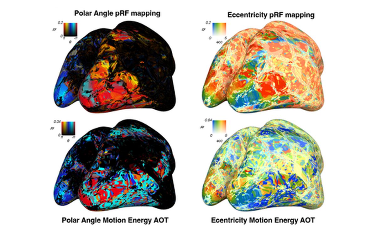
Testing a model voxel by voxel takes many hours of high-quality data from one brain. Other questions need a fast measurement at one location, or timing that fMRI alone cannot give. We design experiments, acquisition methods and analyses for these measurements, and prepare the resulting data for open release.
Approach
With colleagues, we have described the principles of what we call intensive fMRI: extensive sampling of cognitive phenomena in a few individuals, to support computational modeling of single voxels. It depends on experiments designed for a rich hypothesis space and on careful optimization of data quality (Kupers et al., 2024).
Our intensive dataset, Arrow of Time, has about 21 hours of 7T fMRI per participant (1.7 mm isotropic, repetition time 1.6 s), recorded while participants watched 2,160 brief naturalistic videos, plus retinotopy and motion-energy localizers.
Line-scanning fMRI gives up volume coverage to sample a single line through cortex at very high spatial resolution and sampling rate. MEG adds millisecond timing to fMRI’s spatial detail.
What we have found
Line-scanning in humans. Our 7T implementation sampled every 200 ms, and its t-statistics corresponded with those of gradient-echo EPI at the same temporal resolution and voxel volume (R = 0.6 ± 0.2) (Raimondo et al., 2021). Multi-echo readouts and denoising raised temporal signal-to-noise ratio from 4.0 to 36.2, and image-based navigators added motion correction (Raimondo et al., 2022). A spin-echo version gave a sharper line profile, at a cost in functional sensitivity (Raimondo et al., 2023).
Targeting a chosen location. We select a target on each participant’s cortical surface from its anatomy and pRF, place the line there in a second session, and tailor the stimuli to it. For most participants, pRF estimates from the line resembled the target’s; target and best-matching V1 location were on average about 5.5 mm apart (Heij et al., 2023).
Timing from fMRI and MEG. A forward model that predicts MEG responses from each individual’s fMRI visual field maps revealed a processing hierarchy across them: V1 captured most of the variance in the MEG sensors, and did so earlier than extrastriate areas (Eickhoff et al., 2026).
Cortical microstructure in depression. Quantitative 7T MRI of T1 (a proxy for intracortical myelin) and T2* (a proxy for iron) in 48 patients with major depressive disorder and 10 controls linked diagnosis and symptom severity to lower myelination in lateral orbitofrontal cortex, not to iron (Heij et al., 2024).
Body maps from large open datasets. In 7T data from 174 Human Connectome Project participants, a model that maps body-part and visual field tuning at once produced detailed body-part tuning maps from about an hour of rest per participant; during video watching, it revealed an array of somatotopic body maps across the dorsolateral visual system (Hedger et al., 2025). See also Topographic maps.
Open questions
How well do models fit to many hours of data in one brain generalize to new stimuli and to other people? Which questions need a dense, multipurpose dataset, and which a fast measurement aimed at one location? We are preparing Arrow of Time as an open resource; its first release is planned around the turn of the year 2026/27 (get notified).
Papers
8 papers on this theme, selected papers first. Filter the full list.
-
Principles of intensive human neuroimaging
-
Non-invasive mapping of the temporal processing hierarchy in the human visual cortex
-
Quantitative MRI at 7-Tesla reveals novel frontocortical myeloarchitecture anomalies in major depressive disorder
-
A selection and targeting framework of cortical locations for line-scanning fMRI
-
Towards functional spin-echo BOLD line-scanning in humans at 7T
-
Robust high spatio-temporal line-scanning fMRI in humans at 7T using multi-echo readouts, denoising and prospective motion correction
-
A line through the brain: implementation of human line-scanning at 7T for ultra-high spatiotemporal resolution fMRI
-
Porcupine: A visual pipeline tool for neuroimaging analysis
People
Shufan Zhang
PhD student
Former lab members who worked on this theme: Luisa Raimondo, Jurjen Heij.
Software & data
pRFtime
AvailablePython toolbox for spatiotemporal forward modeling of brain signals. It combines pRF maps from fMRI with MEG to estimate when each visual field map contributes to the measured response.
linescanning
AvailableTools for planning, reconstructing and preprocessing line-scanning fMRI data at the Spinoza Centre, including the selection of a target location on the cortical surface.
nsd_access
AvailablePython package for loading data from the Natural Scenes Dataset (NSD), a 7T fMRI dataset: regions of interest, mapping-experiment results and single-trial response estimates, in volume or surface formats.
Arrow of Time
UpcomingAn intensive 7T fMRI dataset of brain responses to 2,160 brief naturalistic videos, with about 21 hours of scanning per participant at 1.7 mm isotropic resolution, plus retinotopy and motion-energy localizers for every participant.
HCP 59k pycortex subject
AvailableA pycortex subject (999999) for the 59k-vertex surfaces of the Human Connectome Project CIFTI format, for plotting surface data on flatmaps.