<?xml version="1.0" encoding="utf-8"?><feed xmlns="http://www.w3.org/2005/Atom" xml:lang="en"><generator uri="https://jekyllrb.com/" version="3.10.0">Jekyll</generator><link href="https://tknapen.github.io/feed.xml" rel="self" type="application/atom+xml" /><link href="https://tknapen.github.io/" rel="alternate" type="text/html" hreflang="en" /><updated>2026-11-10T19:06:07+00:00</updated><id>https://tknapen.github.io/feed.xml</id><title type="html">Knapen Lab</title><subtitle>Knapen Lab, Vrije Universiteit Amsterdam and Spinoza Centre for Neuroimaging: how the brain&apos;s topographic maps organize perception, action and social cognition.</subtitle><author><name>Tomas Knapen</name></author><entry><title type="html">New project on body-based meditation and psilocybin</title><link href="https://tknapen.github.io/2026/11/10/tiny-blue-dot.html" rel="alternate" type="text/html" title="New project on body-based meditation and psilocybin" /><published>2026-11-10T09:00:00+00:00</published><updated>2026-11-10T09:00:00+00:00</updated><id>https://tknapen.github.io/2026/11/10/tiny-blue-dot</id><content type="html" xml:base="https://tknapen.github.io/2026/11/10/tiny-blue-dot.html"><![CDATA[<p>The <a href="https://www.tinybluedotfoundation.org">Tiny Blue Dot Foundation</a> is funding a new project in the lab on body-based meditation and psilocybin. <a href="/people/xaver-funk/">Xaver Funk</a> will join the lab as a postdoctoral researcher in 2027 to work on it.</p>

<p>Related work from the lab includes the body maps we found in visual cortex (<a href="/publications/2025-11-26-NAT/">Hedger et al., 2025</a>) and the effects of psilocybin on visual processing (<a href="/publications/2025-11-21-NC/">Aqil et al., 2025</a>).</p>]]></content><author><name>Tomas Knapen</name></author><summary type="html"><![CDATA[The Tiny Blue Dot Foundation funds a new project in the lab on body-based meditation and psilocybin. Xaver Funk joins as a postdoc in 2027.]]></summary><media:thumbnail xmlns:media="http://search.yahoo.com/mrss/" url="https://tknapen.github.io/assets/img/social-card.png" /><media:content medium="image" url="https://tknapen.github.io/assets/img/social-card.png" xmlns:media="http://search.yahoo.com/mrss/" /></entry><entry><title type="html">Popylar workshop on pRF modeling in Coimbra</title><link href="https://tknapen.github.io/2026/11/06/popylar-workshop-coimbra.html" rel="alternate" type="text/html" title="Popylar workshop on pRF modeling in Coimbra" /><published>2026-11-06T17:00:00+00:00</published><updated>2026-11-06T17:00:00+00:00</updated><id>https://tknapen.github.io/2026/11/06/popylar-workshop-coimbra</id><content type="html" xml:base="https://tknapen.github.io/2026/11/06/popylar-workshop-coimbra.html"><![CDATA[<p>On 5 and 6 November 2026 we ran a two-day workshop on population receptive field (pRF) modeling at the Faculty of Psychology and Educational Sciences of the University of Coimbra, Portugal. The workshop was organized by the Popylar team, on behalf of the Spinoza Centre for Neuroimaging and the Netherlands eScience Center, for PhD students, postdocs and faculty with basic Python skills.</p>

<p>Lectures covered the origins of pRF mapping (Serge Dumoulin), the design of pRF experiments and advanced pRF models (Tomas Knapen) and cognitive pRF models (Ben Harvey). Ilona Bloem presented electrophysiological applications and Marcus Daghlian presented neural contrast sensitivity functions. Malte Lüken of the Netherlands eScience Center introduced the package and discussed modeling choices and best practices.</p>

<p>Most of the time went to hands-on practicals with <a href="https://popylar-org.github.io/prfmodel/">prfmodel</a>, the Python package that the Popylar project develops. Participants fitted Gaussian pRF models to 7T fMRI data, cross-validated and compared models (including difference-of-Gaussians, compressive spatial summation and divisive normalization), fitted connective field models, and applied pRF and related models to electrocorticography, numerosity and contrast sensitivity data. Version 0.2.0 of prfmodel was released for the workshop.</p>

<p>The <a href="https://popylar-org.github.io/2026-11-05_workshop-coimbra/">workshop materials</a> are public: slides from several of the lectures, and the practicals with their solutions, which run in Google Colab. The source is on <a href="https://github.com/popylar-org/2026-11-05_workshop-coimbra">GitHub</a>. To try prfmodel yourself:</p>

<div class="language-bash highlighter-rouge"><div class="highlight"><pre class="highlight"><code>pip <span class="nb">install</span> <span class="s2">"prfmodel[tensorflow]"</span>
</code></pre></div></div>

<p>The <a href="https://popylar-org.github.io/prfmodel/">documentation</a> has tutorials and worked examples. Popylar is supported by the Netherlands eScience Center through its Open eScience Call 2024.</p>]]></content><author><name>Tomas Knapen</name></author><summary type="html"><![CDATA[A two-day hands-on workshop on population receptive field modeling with the new prfmodel Python package, at the University of Coimbra.]]></summary><media:thumbnail xmlns:media="http://search.yahoo.com/mrss/" url="https://tknapen.github.io/assets/img/social-card.png" /><media:content medium="image" url="https://tknapen.github.io/assets/img/social-card.png" xmlns:media="http://search.yahoo.com/mrss/" /></entry><entry><title type="html">PLoS Computational Biology paper combining fMRI and MEG published</title><link href="https://tknapen.github.io/2026/07/10/PCB.html" rel="alternate" type="text/html" title="PLoS Computational Biology paper combining fMRI and MEG published" /><published>2026-07-10T12:00:00+00:00</published><updated>2026-07-10T12:00:00+00:00</updated><id>https://tknapen.github.io/2026/07/10/PCB</id><content type="html" xml:base="https://tknapen.github.io/2026/07/10/PCB.html"><![CDATA[<p><a href="https://doi.org/10.1371/journal.pcbi.1014434">Non-invasive mapping of the temporal processing hierarchy in the human visual cortex</a></p>

<p>K Eickhoff, A Hillebrand, T Knapen, MC de Jong, SO Dumoulin</p>

<p>See the <a href="/publications/2026-07-10-PCB/">publication page</a> for the full reference and links.</p>

<p>The paper introduces a forward model that links population receptive field maps measured with fMRI to responses measured with MEG. For each visual field map, the model predicts its contribution to the MEG signal. Comparing these predictions with the measured responses shows how much each map contributes, and when. Primary visual cortex explained most of the variance in the MEG sensors, and did so earlier than extrastriate areas.</p>

<p>The method is available as the Python toolbox <a href="https://github.com/kateic/pRFtime">pRFtime</a>.</p>]]></content><author><name>Tomas Knapen</name></author><summary type="html"><![CDATA[A forward model that combines pRF maps from fMRI with MEG estimates when each visual field map contributes to the measured response.]]></summary><media:thumbnail xmlns:media="http://search.yahoo.com/mrss/" url="https://tknapen.github.io/assets/img/social-card.png" /><media:content medium="image" url="https://tknapen.github.io/assets/img/social-card.png" xmlns:media="http://search.yahoo.com/mrss/" /></entry><entry><title type="html">Arrow of Time dataset nearing completion</title><link href="https://tknapen.github.io/2026/04/24/AOT.html" rel="alternate" type="text/html" title="Arrow of Time dataset nearing completion" /><published>2026-04-24T12:00:00+00:00</published><updated>2026-04-24T12:00:00+00:00</updated><id>https://tknapen.github.io/2026/04/24/AOT</id><content type="html" xml:base="https://tknapen.github.io/2026/04/24/AOT.html"><![CDATA[<p><strong>Update, November 2026:</strong> The first release of Arrow of Time is planned around the turn of the year. To be notified when it is available, please fill in the <a href="https://forms.gle/BMmfyWEZKL7vU5Va9">sign-up form</a>.</p>

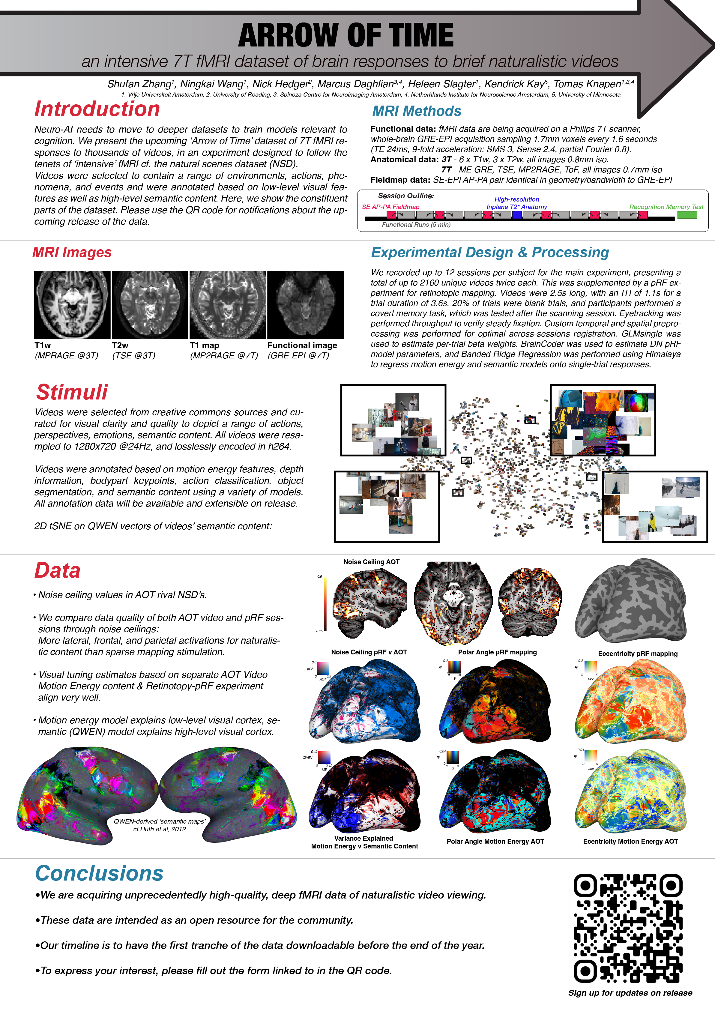
<p>We are in the final stretch of data collection for <strong>Arrow of Time (AOT)</strong>, an intensive 7T fMRI dataset of brain responses to brief naturalistic videos. Over the past year we have been scanning participants on our Philips 7T, and we are now working through the last few subjects on the roster.</p>

<p><img class="col three" src="/img/posts/ccn-AOT.png" alt="Conference poster for the Arrow of Time dataset, with methods, example stimuli, data-quality maps and a QR code linking to the sign-up form." /></p>

<p>Each participant contributes ~21 hours of 7T fMRI at 1.7mm isotropic resolution (TR 1.6 s, SMS 3), viewing 2160 short, creative-commons video clips selected to span a wide range of natural scene and motion statistics. Alongside the main video experiment, every subject also completes separate retinotopy and video motion-energy localizer runs, giving the dataset a rich set of functional anchors per individual.</p>

<p>Early looks at the data are encouraging: noise ceilings across visual cortex are high, the motion-energy and retinotopic maps are crisp, and single-subject semantic structure is already visible in the video-evoked responses. These are exactly the properties we were hoping for when we designed the protocol.</p>

<p>The plan remains to release AOT as an open resource for the community, with a public, downloadable first release (see the update above). If you would like to be notified when the dataset goes live, please fill in <a href="https://forms.gle/BMmfyWEZKL7vU5Va9">the sign-up form</a>. We will keep that list updated as we finalize the release.</p>

<p>More details, including a proper data paper, will follow once scanning wraps.</p>]]></content><author><name>Tomas Knapen</name></author><summary type="html"><![CDATA[Final subjects are being scanned for our intensive 7T fMRI dataset of brain responses to brief naturalistic videos.]]></summary><media:thumbnail xmlns:media="http://search.yahoo.com/mrss/" url="https://tknapen.github.io/assets/img/social-card.png" /><media:content medium="image" url="https://tknapen.github.io/assets/img/social-card.png" xmlns:media="http://search.yahoo.com/mrss/" /></entry><entry><title type="html">Nature paper on vicarious body maps published</title><link href="https://tknapen.github.io/2025/11/26/NAT.html" rel="alternate" type="text/html" title="Nature paper on vicarious body maps published" /><published>2025-11-26T12:00:00+00:00</published><updated>2025-11-26T12:00:00+00:00</updated><id>https://tknapen.github.io/2025/11/26/NAT</id><content type="html" xml:base="https://tknapen.github.io/2025/11/26/NAT.html"><![CDATA[<p>“<a href="https://doi.org/10.1038/s41586-025-09796-0" target="_blank" alt="Vicarious body maps bridge vision and touch in the human brain">Vicarious body maps bridge vision and touch in the human brain</a>”.</p>

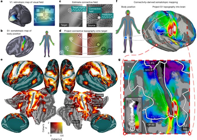
<p>Nicholas Hedger, Thomas Naselaris, Kendrick Kay, Tomas Knapen</p>

<p>See the <a href="/publications/2025-11-26-NAT/">publication page</a> for the full reference and links.</p>

<p><img class="col one right" src="/img/publications/nature_2025_bodymaps.jpg" alt="Overview figure: connective field modeling projects the visual field map of V1 and the body map of S1 onto the rest of cortex, revealing somatotopic maps on inflated and flattened cortical surfaces." /></p>

<p><strong>Abstract</strong> Our sensory systems work together to generate a cohesive experience of the world. Observing touch to others activates regions representing one’s own touch, suggesting that visual processing recruits touch-related computations. To examine this interface, we developed a model that jointly maps somatosensory body-part tuning and visual field tuning across the brain. During rest, the model recovered detailed body-part tuning in endogenous somatotopic networks. During movie viewing, somatotopic tuning explained responses throughout dorsolateral visual cortex, revealing multiple somatotopic body maps tiled across cortex. Body-position tuning aligned with visual tuning, predicting both visual field preferences and category preferences for body parts. These results suggest aligned visual-somatosensory topographic maps that connect visual and bodily reference frames and help translate sensory input into representations useful for action, social cognition, and semantic processing.</p>]]></content><author><name>Tomas Knapen</name></author><summary type="html"><![CDATA[Vicarious body maps link visual and tactile processing in human cortex.]]></summary><media:thumbnail xmlns:media="http://search.yahoo.com/mrss/" url="https://tknapen.github.io/assets/img/social-card.png" /><media:content medium="image" url="https://tknapen.github.io/assets/img/social-card.png" xmlns:media="http://search.yahoo.com/mrss/" /></entry><entry><title type="html">Nature Communications paper on psilocybin and visual context published</title><link href="https://tknapen.github.io/2025/11/21/NC.html" rel="alternate" type="text/html" title="Nature Communications paper on psilocybin and visual context published" /><published>2025-11-21T12:00:00+00:00</published><updated>2025-11-21T12:00:00+00:00</updated><id>https://tknapen.github.io/2025/11/21/NC</id><content type="html" xml:base="https://tknapen.github.io/2025/11/21/NC.html"><![CDATA[<p>“<a href="https://doi.org/10.1038/s41467-025-65150-y" target="_blank" alt="Psilocybin alters visual contextual computations">Psilocybin alters visual contextual computations</a>”.</p>

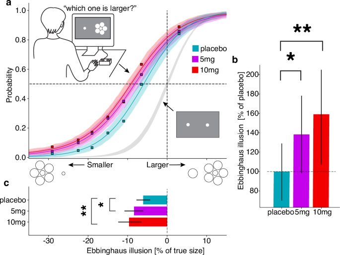
<p>Marco Aqil, Gilles de Hollander, Nina Vreugdenhil, Tomas Knapen, Serge O. Dumoulin</p>

<p>See the <a href="/publications/2025-11-21-NC/">publication page</a> for the full reference and links.</p>

<p><img class="col one right" src="/img/publications/natcomms_2025_psilocybin.png" alt="Psychometric curves and bar plots showing a larger Ebbinghaus illusion after 5 mg and 10 mg psilocybin than after placebo." /></p>

<p><strong>Abstract</strong> Psilocybin alters perception and brain dynamics. Here we combined psychophysics, ultra-high-field fMRI, and computational modeling to test its effects on contextual computations. We found that psilocybin alters contextual perception in the Ebbinghaus illusion, alters contextual modulation in cortical responses to visual stimuli, and is captured by a computational model linking these effects. These findings suggest altered contextual computation as a potential general mechanism underlying psychedelic action.</p>]]></content><author><name>Tomas Knapen</name></author><summary type="html"><![CDATA[Effects of psilocybin on contextual computations in visual processing.]]></summary><media:thumbnail xmlns:media="http://search.yahoo.com/mrss/" url="https://tknapen.github.io/assets/img/social-card.png" /><media:content medium="image" url="https://tknapen.github.io/assets/img/social-card.png" xmlns:media="http://search.yahoo.com/mrss/" /></entry><entry><title type="html">Nature Human Behaviour paper on binocular rivalry published</title><link href="https://tknapen.github.io/2025/11/13/NHB.html" rel="alternate" type="text/html" title="Nature Human Behaviour paper on binocular rivalry published" /><published>2025-11-13T12:00:00+00:00</published><updated>2025-11-13T12:00:00+00:00</updated><id>https://tknapen.github.io/2025/11/13/NHB</id><content type="html" xml:base="https://tknapen.github.io/2025/11/13/NHB.html"><![CDATA[<p>“<a href="https://doi.org/10.1038/s41562-025-02320-4" target="_blank" alt="Mesoscale cortical mechanisms of perceptual conflict resolution in binocular rivalry">Mesoscale cortical mechanisms of perceptual conflict resolution in binocular rivalry</a>”.</p>

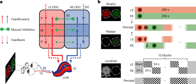
<p>Chencan Qian, Zihao Zhang, Zhiqiang Chen, Gilles de Hollander, Tomas Knapen, Sheng He, Peng Zhang</p>

<p>See the <a href="/publications/2025-11-13-NHB/">publication page</a> for the full reference and links.</p>

<p><img class="col one right" src="/img/publications/nhb_2025_binocular_rivalry.png" alt="Schematic of the binocular rivalry study: feedforward input, mutual inhibition and feedback between left-eye and right-eye ocular dominance columns across cortical depth, with timelines of the rivalry, replay and localizer conditions." /></p>

<p><strong>Abstract</strong> How does the human brain resolve conflicts in sensory input to generate conscious perception? Using high-resolution 7 T functional MRI, we investigated column- and layer-specific activity in cortical and subcortical regions during binocular rivalry. Eye-specific rivalry arose from interocular inhibition between adjacent ocular dominance columns in superficial primary visual cortex, but not between ocular layers in the lateral geniculate nucleus. Eye-specific feedback from intraparietal sulcus actively biased and synchronized local competitions in primary visual cortex into coherent perceptual representations, even without awareness of eye-of-origin information. These findings identify mesoscale mechanisms of perceptual conflict resolution in humans: local conflicts are resolved by inhibitory sensory-cortical microcircuits, while parietal feedback biases and integrates local competition into unified conscious perception.</p>]]></content><author><name>Tomas Knapen</name></author><summary type="html"><![CDATA[Mesoscale mechanisms of perceptual conflict resolution in binocular rivalry.]]></summary><media:thumbnail xmlns:media="http://search.yahoo.com/mrss/" url="https://tknapen.github.io/assets/img/social-card.png" /><media:content medium="image" url="https://tknapen.github.io/assets/img/social-card.png" xmlns:media="http://search.yahoo.com/mrss/" /></entry><entry><title type="html">Nature Neuroscience perspective on consciousness theory published</title><link href="https://tknapen.github.io/2025/03/10/NN.html" rel="alternate" type="text/html" title="Nature Neuroscience perspective on consciousness theory published" /><published>2025-03-10T12:00:00+00:00</published><updated>2025-03-10T12:00:00+00:00</updated><id>https://tknapen.github.io/2025/03/10/NN</id><content type="html" xml:base="https://tknapen.github.io/2025/03/10/NN.html"><![CDATA[<p>“<a href="https://doi.org/10.1038/s41593-025-01881-x" target="_blank" alt="What makes a theory of consciousness unscientific?">What makes a theory of consciousness unscientific?</a>”.</p>

<p>Michal Klincewicz et al. (including Tomas Knapen)</p>

<p>See the <a href="/publications/2025-03-10-NN/">publication page</a> for the full reference and links.</p>

<p><strong>Abstract</strong> Theories of consciousness have a long and controversial history. One well-known proposal, integrated information theory, has recently been labeled as pseudoscience, which has caused a heated open debate. Here we discuss the case and argue that the theory is unscientific because its core claims are untestable even in principle.</p>]]></content><author><name>Tomas Knapen</name></author><summary type="html"><![CDATA[Perspective on scientific standards for theories of consciousness.]]></summary><media:thumbnail xmlns:media="http://search.yahoo.com/mrss/" url="https://tknapen.github.io/assets/img/social-card.png" /><media:content medium="image" url="https://tknapen.github.io/assets/img/social-card.png" xmlns:media="http://search.yahoo.com/mrss/" /></entry><entry><title type="html">Translational Psychiatry paper on 7T myeloarchitecture published</title><link href="https://tknapen.github.io/2024/06/20/TP.html" rel="alternate" type="text/html" title="Translational Psychiatry paper on 7T myeloarchitecture published" /><published>2024-06-20T12:00:00+00:00</published><updated>2024-06-20T12:00:00+00:00</updated><id>https://tknapen.github.io/2024/06/20/TP</id><content type="html" xml:base="https://tknapen.github.io/2024/06/20/TP.html"><![CDATA[<p>“<a href="https://doi.org/10.1038/s41398-024-02976-y" target="_blank" alt="Quantitative MRI at 7-Tesla reveals novel frontocortical myeloarchitecture anomalies in major depressive disorder">Quantitative MRI at 7-Tesla reveals novel frontocortical myeloarchitecture anomalies in major depressive disorder</a>”.</p>

<p>Jurjen Heij, Wietske van der Zwaag, Tomas Knapen, Matthan W.A. Caan, Birte Forstman, Dick J. Veltman, Guido van Wingen, Moji Aghajani</p>

<p>See the <a href="/publications/2024-06-20-TP/">publication page</a> for the full reference and links.</p>

<p><img class="col one right" src="/img/publications/transpsy_2024_myeloarchitecture.png" alt="Cortical maps of T1 and T2* relaxation times, and box plots of left and right hemisphere values in rostral anterior cingulate, lateral orbitofrontal and medial orbitofrontal cortex." /></p>

<p><strong>Abstract</strong> Whereas meta-analytical data highlight abnormal frontocortical macrostructure (thickness/surface area/volume) in Major Depressive Disorder (MDD), the underlying microstructural processes remain uncharted, due to the use of conventional MRI scanners and acquisition techniques. We uniquely combined Ultra-High Field MRI at 7.0 Tesla with Quantitative Imaging to map intracortical myelin (proxied by longitudinal relaxation time T<sub>1</sub>) and iron concentration (proxied by transverse relaxation time T<sub>2</sub>*), microstructural processes deemed particularly germane to cortical macrostructure. Informed by meta-analytical evidence, we focused specifically on orbitofrontal and rostral anterior cingulate cortices among adult MDD patients (N = 48) and matched healthy controls (HC; N = 10). Analyses probed the association of MDD diagnosis and clinical profile (severity, medication use, comorbid anxiety disorders, childhood trauma) with aforementioned microstructural properties. MDD diagnosis (p’s &lt; 0.05, Cohen’s D = 0.55-0.66) and symptom severity (p’s &lt; 0.01, r = 0.271-0.267) both related to decreased intracortical myelination (higher T<sub>1</sub> values) within the lateral orbitofrontal cortex, a region tightly coupled to processing negative affect and feelings of sadness in MDD. No relations were found with local iron concentrations. These findings allow uniquely fine-grained insights on frontocortical microstructure in MDD, and cautiously point to intracortical demyelination as a possible driver of macroscale cortical disintegrity in MDD.</p>]]></content><author><name>Tomas Knapen</name></author><summary type="html"><![CDATA[Quantitative 7T MRI findings on frontocortical myeloarchitecture in depression.]]></summary><media:thumbnail xmlns:media="http://search.yahoo.com/mrss/" url="https://tknapen.github.io/assets/img/social-card.png" /><media:content medium="image" url="https://tknapen.github.io/assets/img/social-card.png" xmlns:media="http://search.yahoo.com/mrss/" /></entry><entry><title type="html">Review on visuospatial coding in Trends in Cognitive Sciences</title><link href="https://tknapen.github.io/2021/11/16/TiCS.html" rel="alternate" type="text/html" title="Review on visuospatial coding in Trends in Cognitive Sciences" /><published>2021-11-16T12:00:00+00:00</published><updated>2021-11-16T12:00:00+00:00</updated><id>https://tknapen.github.io/2021/11/16/TiCS</id><content type="html" xml:base="https://tknapen.github.io/2021/11/16/TiCS.html"><![CDATA[<p>“<a href="https://doi.org/10.1016/j.tics.2021.10.011" target="_blank" alt="Visuospatial coding as ubiquitous scaffolding for human cognition">Visuospatial coding as ubiquitous scaffolding for human cognition</a>”.</p>

<p>I Groen, T Dekker, T Knapen, E Silson</p>

<p>See the <a href="/publications/2021-11-16-TiCS/">publication page</a> for the full reference and links.</p>

<p><strong><em>Highlights</em></strong></p>

<ul>
  <li>Visuospatial coding in the brain comes in at least two forms: retinotopic maps and visual field biases. Visuospatial coding is not restricted to early visual cortex but extends throughout the brain.</li>
  <li>Evidence for visuospatial coding is present across a broad array of functionally disparate systems beyond visual cortex including the cerebellum, hippocampus, frontal eye fields, and the default mode network.</li>
  <li>Category-selective regions of visual cortex often spatially overlap multiple retinotopic maps, and exhibit biases for visual field locations typically containing their preferred stimulus.</li>
  <li>Visuospatial coding is present prior to the emergence of category selectivity and offers a plausible explanation for why such regions emerge where they do in cortex. Visual location tuning of these regions changes throughout development and correlates with changes in viewing behaviour.</li>
  <li>The ubiquity of visuospatial coding suggests that the brain does not ‘throw away’ this frame of reference but instead holds onto it to scaffold human cognition in a common framework.</li>
</ul>

<p><strong>Abstract</strong> For more than 100 years we have known that the visual field is mapped onto the surface of visual cortex, imposing an inherently spatial reference frame on visual information processing. Recent studies highlight visuospatial coding not only throughout visual cortex, but also brain areas not typically considered visual. Such widespread access to visuospatial coding raises important questions about its role in wider cognitive functioning. Here, we synthesise these recent developments and propose that visuospatial coding scaffolds human cognition by providing a reference frame through which neural computations interface with environmental statistics and task demands via perception–action loops.</p>]]></content><author><name>Tomas Knapen</name></author><summary type="html"><![CDATA[Review on how the brain uses visual space as a fundamental coding mechanism]]></summary><media:thumbnail xmlns:media="http://search.yahoo.com/mrss/" url="https://tknapen.github.io/assets/img/social-card.png" /><media:content medium="image" url="https://tknapen.github.io/assets/img/social-card.png" xmlns:media="http://search.yahoo.com/mrss/" /></entry><entry><title type="html">PNAS paper on divisive normalization in the visual hierarchy</title><link href="https://tknapen.github.io/2021/11/15/PNAS.html" rel="alternate" type="text/html" title="PNAS paper on divisive normalization in the visual hierarchy" /><published>2021-11-15T12:00:00+00:00</published><updated>2021-11-15T12:00:00+00:00</updated><id>https://tknapen.github.io/2021/11/15/PNAS</id><content type="html" xml:base="https://tknapen.github.io/2021/11/15/PNAS.html"><![CDATA[<p>“<a href="https://doi.org/10.1073/pnas.2108713118" target="_blank" alt="Divisive normalization unifies disparate response signatures throughout the human visual hierarchy">Divisive normalization unifies disparate response signatures throughout the human visual hierarchy</a>”.</p>

<p>M Aqil, T Knapen, S Dumoulin</p>

<p>See the <a href="/publications/2021-11-15-PNAS/">publication page</a> for the full reference and links.</p>

<p><strong><em>SIGNIFICANCE STATEMENT</em></strong></p>

<p>A canonical neural computation is a mathematical operation applied by the brain in a wide variety of contexts and capable of explaining and unifying seemingly unrelated neural and perceptual phenomena. Here, we use a combination of state-of-the-art experiments (ultra-high-field functional MRI) and mathematical methods (population receptive field [pRF] modeling) to uniquely demonstrate the role of divisive normalization (DN) as the canonical neural computation underlying visuospatial responses throughout the human visual hierarchy. The DN pRF model provides a tool to investigate and interpret the computational processes underlying neural responses in human and animal recordings, but also in clinical and cognitive dimensions.</p>

<p><strong>Abstract</strong> Neural processing is hypothesized to apply the same mathematical operations in a variety of contexts, implementing so-called canonical neural computations. Divisive normalization (DN) is considered a prime candidate for a canonical computation. Here, we propose a population receptive field (pRF) model based on DN and evaluate it using ultra-high-field functional MRI (fMRI). The DN model parsimoniously captures seemingly disparate response signatures with a single computation, superseding existing pRF models in both performance and biological plausibility. We observe systematic variations in specific DN model parameters across the visual hierarchy and show how they relate to differences in response modulation and visuospatial information integration. The DN model delivers a unifying framework for visuospatial responses throughout the human visual hierarchy and provides insights into its underlying information-encoding computations. These findings extend the role of DN as a canonical computation to neuronal populations throughout the human visual hierarchy.</p>]]></content><author><name>Tomas Knapen</name></author><summary type="html"><![CDATA[Our new divisive normalization population receptive field model explains responses throughout visual cortex.]]></summary><media:thumbnail xmlns:media="http://search.yahoo.com/mrss/" url="https://tknapen.github.io/assets/img/social-card.png" /><media:content medium="image" url="https://tknapen.github.io/assets/img/social-card.png" xmlns:media="http://search.yahoo.com/mrss/" /></entry><entry><title type="html">Welcome to Ron, Ningkai and Niklas</title><link href="https://tknapen.github.io/2021/09/01/people.html" rel="alternate" type="text/html" title="Welcome to Ron, Ningkai and Niklas" /><published>2021-09-01T12:00:00+00:00</published><updated>2021-09-01T12:00:00+00:00</updated><id>https://tknapen.github.io/2021/09/01/people</id><content type="html" xml:base="https://tknapen.github.io/2021/09/01/people.html"><![CDATA[<p><a href="/people/2021-09-01-rk/">Ron</a> &amp; <a href="/people/2021-09-02-nw/">Ningkai</a> are new PhD students in the lab. Niklas is a new Master’s student from AI, that’s also going to be lab assistant in the Dumoulin lab at Spinoza.</p>]]></content><author><name>Tomas Knapen</name></author><summary type="html"><![CDATA[New PhD and master's students starting this month.]]></summary><media:thumbnail xmlns:media="http://search.yahoo.com/mrss/" url="https://tknapen.github.io/assets/img/social-card.png" /><media:content medium="image" url="https://tknapen.github.io/assets/img/social-card.png" xmlns:media="http://search.yahoo.com/mrss/" /></entry><entry><title type="html">Line-scanning fMRI at 7T</title><link href="https://tknapen.github.io/2021/08/20/JCBFM.html" rel="alternate" type="text/html" title="Line-scanning fMRI at 7T" /><published>2021-08-20T12:00:00+00:00</published><updated>2021-08-20T12:00:00+00:00</updated><id>https://tknapen.github.io/2021/08/20/JCBFM</id><content type="html" xml:base="https://tknapen.github.io/2021/08/20/JCBFM.html"><![CDATA[<p>“<a href="https://doi.org/10.1177/0271678X211037266" target="_blank" alt="A line through the brain: implementation of human line-scanning at 7T for ultra-high spatiotemporal resolution fMRI">A line through the brain: implementation of human line-scanning at 7T for ultra-high spatiotemporal resolution fMRI</a>”.</p>

<p>Luisa Raimondo, Tomas Knapen, ĺcaro AF Oliveira, Xin Yu, Serge O Dumoulin, Wietske van der Zwaag, Jeroen CW Siero</p>

<p>See the <a href="/publications/2021-08-20-JCBFM/">publication page</a> for the full reference and links.</p>

<p><strong>Abstract</strong> Functional magnetic resonance imaging (fMRI) is a widely used tool in neuroscience to detect neurally evoked responses, e.g. the blood oxygenation level-dependent (BOLD) signal. Typically, BOLD fMRI has millimeter spatial resolution and temporal resolution of one to few seconds. To study the sub-millimeter structures and activity of the cortical gray matter, the field needs an fMRI method with high spatial and temporal resolution. Line-scanning fMRI achieves very high spatial resolution and high sampling rate, at the cost of a sacrifice in volume coverage. Here, we present a human line-scanning implementation on a 7T MRI system. First, we investigate the quality of the saturation pulses that suppress MR signal outside the line. Second, we established the best coil combination for reconstruction. Finally, we applied the line-scanning method in the occipital lobe during a visual stimulation task, showing BOLD responses along cortical depth, every 250 µm with a 200 ms repetition time (TR). We found a good correspondence of t-statistics values with 2D gradient-echo echo planar imaging (GE-EPI) BOLD fMRI data with the same temporal resolution and voxel volume (R = 0.6 ± 0.2). In summary, we demonstrate the feasibility of line-scanning in humans and this opens line-scanning fMRI for applications in cognitive and clinical neuroscience.</p>]]></content><author><name>Tomas Knapen</name></author><summary type="html"><![CDATA[Line-scanning at 7T samples BOLD responses across cortical depth every 250 µm with a 200 ms repetition time.]]></summary><media:thumbnail xmlns:media="http://search.yahoo.com/mrss/" url="https://tknapen.github.io/assets/img/social-card.png" /><media:content medium="image" url="https://tknapen.github.io/assets/img/social-card.png" xmlns:media="http://search.yahoo.com/mrss/" /></entry><entry><title type="html">PNAS paper on retinotopic processing in the default network and hippocampus</title><link href="https://tknapen.github.io/2021/01/12/PNAS.html" rel="alternate" type="text/html" title="PNAS paper on retinotopic processing in the default network and hippocampus" /><published>2021-01-12T12:00:00+00:00</published><updated>2021-01-12T12:00:00+00:00</updated><id>https://tknapen.github.io/2021/01/12/PNAS</id><content type="html" xml:base="https://tknapen.github.io/2021/01/12/PNAS.html"><![CDATA[<p>“<a href="https://doi.org/10.1073/pnas.2017032118" target="_blank" alt="Topographic connectivity reveals task-dependent retinotopic processing throughout the human brain">Topographic connectivity reveals task-dependent retinotopic processing throughout the human brain</a>”.</p>

<p>T Knapen</p>

<p>See the <a href="/publications/2021-01-12-PNAS/">publication page</a> for the full reference and links.</p>

<p><img class="col two right" src="/img/science/999999/999999-RS-movie.png" alt="Flattened maps of both cerebral hemispheres comparing topographic connectivity during rest and movie watching, with major sulci labeled." /></p>

<p><strong>Abstract</strong> The human visual system is organized as a hierarchy of maps that share the topography of the retina. Known retinotopic maps have been identified using simple visual stimuli under strict fixation, conditions different from everyday vision which is active, dynamic, and complex. This means that it remains unknown how much of the brain is truly visually organized. Here I demonstrate widespread stable visual organization beyond the traditional visual system, in default-mode network and hippocampus. Detailed topographic connectivity with primary visual cortex during movie-watching, resting-state, and retinotopic-mapping experiments revealed that visual–spatial representations throughout the brain are warped by cognitive state. Specifically, traditionally visual regions alternate with default-mode network and hippocampus in preferentially representing the center of the visual field. This visual role of default-mode network and hippocampus would allow these regions to interface between abstract memories and concrete sensory impressions. Together, these results indicate that visual–spatial organization is a fundamental coding principle that structures the communication between distant brain regions.</p>]]></content><author><name>Tomas Knapen</name></author><summary type="html"><![CDATA[Using topographic connectivity mapping, we show that hippocampus is visually organized.]]></summary><media:thumbnail xmlns:media="http://search.yahoo.com/mrss/" url="https://tknapen.github.io/assets/img/social-card.png" /><media:content medium="image" url="https://tknapen.github.io/assets/img/social-card.png" xmlns:media="http://search.yahoo.com/mrss/" /></entry><entry><title type="html">Preprint on the visual organization of the default network and hippocampus</title><link href="https://tknapen.github.io/2020/08/08/biorxiv.html" rel="alternate" type="text/html" title="Preprint on the visual organization of the default network and hippocampus" /><published>2020-08-08T12:00:00+00:00</published><updated>2020-08-08T12:00:00+00:00</updated><id>https://tknapen.github.io/2020/08/08/biorxiv</id><content type="html" xml:base="https://tknapen.github.io/2020/08/08/biorxiv.html"><![CDATA[<p>“<a href="https://www.biorxiv.org/content/10.1101/2020.07.30.228403v1" target="_blank" alt="Topographic connectivity reveals task-dependent retinotopic processing throughout the human brain">Topographic connectivity reveals task-dependent retinotopic processing throughout the human brain</a>”.</p>

<p>T Knapen</p>

<p>The published version is on the <a href="/publications/2021-01-12-PNAS/">publication page</a>.</p>

<p><strong>New preprint online @ biorxiv:</strong> The human visual system is organized as a hierarchy of maps that share the topography of the retina. These retinotopic maps have been identified throughout the brain, but how much of the brain is visually organized remains unknown. Here we demonstrate widespread stable visual organization beyond the traditional visual system. We analyzed detailed topographic connectivity with primary visual cortex during moviewatching, rest, and retinotopic mapping experiments to reveal that visual-spatial representations are warped by experimental condition and cognitive state. Specifically, traditionally visual regions alternate with default mode network and hippocampus in preferentially representing the center of the visual field. This visual role of hippocampus would allow it to implement sensory predictions by interfacing between abstract memories and concrete perceptions. These results indicate that pervasive sensory coding facilitates the communication between far-flung brain regions.</p>]]></content><author><name>Tomas Knapen</name></author><summary type="html"><![CDATA[Using topographic connectivity mapping, we show that hippocampus is visually organized.]]></summary><media:thumbnail xmlns:media="http://search.yahoo.com/mrss/" url="https://tknapen.github.io/assets/img/social-card.png" /><media:content medium="image" url="https://tknapen.github.io/assets/img/social-card.png" xmlns:media="http://search.yahoo.com/mrss/" /></entry><entry><title type="html">Current Biology paper on intracranial recordings during binocular rivalry</title><link href="https://tknapen.github.io/2020/07/02/CB.html" rel="alternate" type="text/html" title="Current Biology paper on intracranial recordings during binocular rivalry" /><published>2020-07-02T12:00:00+00:00</published><updated>2020-07-02T12:00:00+00:00</updated><id>https://tknapen.github.io/2020/07/02/CB</id><content type="html" xml:base="https://tknapen.github.io/2020/07/02/CB.html"><![CDATA[<p>“<a href="https://www.cell.com/current-biology/fulltext/S0960-9822(20)30769-7" target="_blank" alt="Intracranial Recordings Reveal Unique Shape and Timing of Responses in Human Visual Cortex during Illusory Visual Events">Intracranial Recordings Reveal Unique Shape and Timing of Responses in Human Visual Cortex during Illusory Visual Events</a>”.</p>

<p>Maartje C. de Jong, Mariska J. Vansteensel, Raymond van Ee, Frans S.S. Leijten, Nick F. Ramsey, H. Chris Dijkerman, Serge O. Dumoulin &amp; Tomas Knapen</p>

<p>See the <a href="/publications/2020-08-17-CB/">publication page</a> for the full reference and links.</p>

<p><img class="col two right" src="/img/posts/duckrabbit.png" alt="A rabbit's head with a duck's bill, an object that can be seen as either animal." /></p>

<p><strong>New paper online @ Current Biology:</strong> During binocular rivalry, perception spontaneously changes without any alteration to the visual stimulus. What neural events bring about this illusion that a constant stimulus is changing? We recorded from intracranial electrodes placed on the occipital and posterior temporal cortex of two patients with epilepsy while they experienced illusory changes of a face-house binocular-rivalry stimulus or observed a control stimulus that physically changed. We performed within-patient comparisons of broadband high-frequency responses, focusing on single epochs recorded along the ventral processing stream. We found transient face- and house-selective responses localized to the same electrodes for illusory and physical changes, but the temporal characteristics of these responses markedly differed. In comparison with physical changes, responses to illusory changes were longer lasting, in particular exhibiting a characteristic slow rise. Furthermore, the temporal order of responses across the visual hierarchy was reversed for illusory as compared to physical changes: for illusory changes, higher order fusiform and parahippocampal regions responded before lower order occipital regions. Our tentative interpretation of these findings is that two stages underlie the initiation of illusory changes: a destabilization stage in which activity associated with the impending change gradually accumulates across the visual hierarchy, ultimately graduating in a top-down cascade of activity that may stabilize the new perceptual interpretation of the stimulus.</p>]]></content><author><name>Tomas Knapen</name></author><summary type="html"><![CDATA[Intracranial recordings reveal unique shape and timing of responses in human visual cortex during illusory visual events.]]></summary><media:thumbnail xmlns:media="http://search.yahoo.com/mrss/" url="https://tknapen.github.io/assets/img/social-card.png" /><media:content medium="image" url="https://tknapen.github.io/assets/img/social-card.png" xmlns:media="http://search.yahoo.com/mrss/" /></entry><entry><title type="html">Preprint on visual cortex in reinforcement learning</title><link href="https://tknapen.github.io/2019/01/22/biorxiv.html" rel="alternate" type="text/html" title="Preprint on visual cortex in reinforcement learning" /><published>2019-01-22T12:00:00+00:00</published><updated>2019-01-22T12:00:00+00:00</updated><id>https://tknapen.github.io/2019/01/22/biorxiv</id><content type="html" xml:base="https://tknapen.github.io/2019/01/22/biorxiv.html"><![CDATA[<p>“<a href="https://www.biorxiv.org/content/early/2019/01/21/523340" target="_blank" alt="Learning in visual regions as support for the bias in future value-driven choice">Learning in visual regions as support for the bias in future value-driven choice</a>”.</p>

<p>S Jahfari, J Theeuwes, T Knapen</p>

<p>The published version is on the <a href="/publications/2020-04-01-CC/">publication page</a>.</p>

<p><img class="col two right" src="/img/prf/retmaps.png" alt="Inflated cortical surfaces colored by preferred polar angle, with visual areas from V1 to the intraparietal sulcus outlined and labeled." /></p>

<p><strong>New preprint online @ biorxiv:</strong> Learning biases decision-making towards higher expected outcomes. Cognitive theories describe this through the tracking of value and outcome evaluations within striatum and prefrontal cortex. Decisions however first require processing of sensory input, and to-date, far less is known about the learning perception interplay. This fMRI study (N=43), relates visual BOLD responses to value-beliefs during choice, and, signed prediction errors after outcomes. To understand these relationships, which co-occurred in striatum, we next evaluated relevance with the prediction of future value-based choices, using a separate transfer-phase with learning already established. We decoded choice outcomes with a 69% accuracy with a machine learning algorithm that was given trial-by-trial BOLD from visual regions alongside traditional prefrontal, and striatal regions. Importantly, this decoding of value-driven choice outcomes again showed an important role for visual activity. These results raise the intriguing possibility that value learning in visual cortex is supportive for the striatal bias towards valued options.</p>]]></content><author><name>Tomas Knapen</name></author><summary type="html"><![CDATA[Learning in visual regions as support for the bias in future value-driven choice]]></summary><media:thumbnail xmlns:media="http://search.yahoo.com/mrss/" url="https://tknapen.github.io/assets/img/social-card.png" /><media:content medium="image" url="https://tknapen.github.io/assets/img/social-card.png" xmlns:media="http://search.yahoo.com/mrss/" /></entry><entry><title type="html">PLoS ONE paper on saccadic adaptation published</title><link href="https://tknapen.github.io/2019/01/16/PONE.html" rel="alternate" type="text/html" title="PLoS ONE paper on saccadic adaptation published" /><published>2019-01-16T12:00:00+00:00</published><updated>2019-01-16T12:00:00+00:00</updated><id>https://tknapen.github.io/2019/01/16/PONE</id><content type="html" xml:base="https://tknapen.github.io/2019/01/16/PONE.html"><![CDATA[<p>“<a href="https://journals.plos.org/plosone/article?id=10.1371/journal.pone.0203248" target="_blank" alt="Implicit and explicit learning in reactive and voluntary saccade adaptation">Implicit and explicit learning in reactive and voluntary saccade adaptation</a>”.</p>

<p>D.M. van Es, T. Knapen</p>

<p>See the <a href="/publications/2019-01-16-PO/">publication page</a> for the full reference and links.</p>

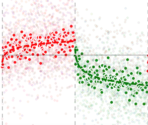
<p><img class="col two right" src="/img/science/sa.png" alt="Saccade adaptation data over trials, with an increasing phase in red and a decreasing phase in green, each with a fitted learning curve." /></p>

<p><strong>New paper accepted in PLoS ONE:</strong> Saccades can either be elicited automatically by salient peripheral stimuli or can additionally depend on explicit cognitive goals. Similarly, it is thought that motor adaptation is driven by the combination of a more automatic, implicit process and a more explicit, cognitive process. However, the degree to which such implicit and explicit learning contribute to the adaptation of more reactive and voluntary saccades remains elusive. To study this question, we employed a global saccadic adaptation paradigm with both increasing and decreasing saccade amplitudes. We assessed the resulting adaptation using a dual state model of motor adaptation. This model decomposes learning into a fast and slow process, which are thought to constitute explicit and implicit learning, respectively. Our results show that adaptation of reactive saccades is equally driven by fast and slow learning, while fast learning is nearly absent when adapting voluntary (i.e. scanning) saccades. This pattern of results was present both when saccade gain was increased or decreased. Our results suggest that the increased cognitive demands associated with voluntary compared to reactive saccade planning interfere specifically with explicit learning.</p>]]></content><author><name>Tomas Knapen</name></author><summary type="html"><![CDATA[Implicit and explicit learning in reactive and voluntary saccade adaptation]]></summary><media:thumbnail xmlns:media="http://search.yahoo.com/mrss/" url="https://tknapen.github.io/assets/img/social-card.png" /><media:content medium="image" url="https://tknapen.github.io/assets/img/social-card.png" xmlns:media="http://search.yahoo.com/mrss/" /></entry><entry><title type="html">eLife paper on attentional warping of visual space published</title><link href="https://tknapen.github.io/2018/12/07/eLife.html" rel="alternate" type="text/html" title="eLife paper on attentional warping of visual space published" /><published>2018-12-07T12:00:00+00:00</published><updated>2018-12-07T12:00:00+00:00</updated><id>https://tknapen.github.io/2018/12/07/eLife</id><content type="html" xml:base="https://tknapen.github.io/2018/12/07/eLife.html"><![CDATA[<p>“<a href="https://elifesciences.org/articles/36928" target="_blank" alt="Spatial sampling in human visual cortex is modulated by both spatial and feature-based attention">Spatial sampling in human visual cortex is modulated by both spatial and feature-based attention</a>”.</p>

<p>D.M. van Es, J. Theeuwes, T. Knapen</p>

<p>See the <a href="/publications/2018-12-07-EL/">publication page</a> for the full reference and links.</p>

<p><img class="col two right" src="/img/prf/retmaps.png" alt="Inflated cortical surfaces colored by preferred polar angle, with visual areas from V1 to the intraparietal sulcus outlined and labeled." /></p>

<p><strong>New paper published in eLife:</strong> Spatial attention changes the sampling of visual space. Behavioral studies suggest that feature-based attention modulates this resampling to optimize the attended feature’s sampling. We investigate this hypothesis by estimating spatial sampling in visual cortex while independently varying both feature-based and spatial attention. Our results show that spatial and feature-based attention interacted: resampling of visual space depended on both the attended location and feature (color vs. temporal frequency). This interaction occurred similarly throughout visual cortex, regardless of an area’s overall feature preference. However, the interaction did depend on spatial sampling properties of voxels that prefer the attended feature. These findings are parsimoniously explained by variations in the precision of an attentional gain field. Our results demonstrate that the deployment of spatial attention is tailored to the spatial sampling properties of units that are sensitive to the attended feature.</p>]]></content><author><name>Tomas Knapen</name></author><summary type="html"><![CDATA[Spatial sampling in human visual cortex is modulated by both spatial and feature-based attention]]></summary><media:thumbnail xmlns:media="http://search.yahoo.com/mrss/" url="https://tknapen.github.io/assets/img/social-card.png" /><media:content medium="image" url="https://tknapen.github.io/assets/img/social-card.png" xmlns:media="http://search.yahoo.com/mrss/" /></entry><entry><title type="html">Preprint on retinotopic maps in the cerebellum</title><link href="https://tknapen.github.io/2018/10/29/biorxiv.html" rel="alternate" type="text/html" title="Preprint on retinotopic maps in the cerebellum" /><published>2018-10-29T12:00:00+00:00</published><updated>2018-10-29T12:00:00+00:00</updated><id>https://tknapen.github.io/2018/10/29/biorxiv</id><content type="html" xml:base="https://tknapen.github.io/2018/10/29/biorxiv.html"><![CDATA[<p>“<a href="https://www.biorxiv.org/content/early/2018/10/29/455170" target="_blank" alt="Retinotopic maps of visual space in the human cerebellum">Retinotopic maps of visual space in the human cerebellum</a>”.</p>

<p>D.M. van Es, W. van der Zwaag, T. Knapen</p>

<p>The published version is on the <a href="/publications/2019-05-20-CB/">publication page</a>.</p>

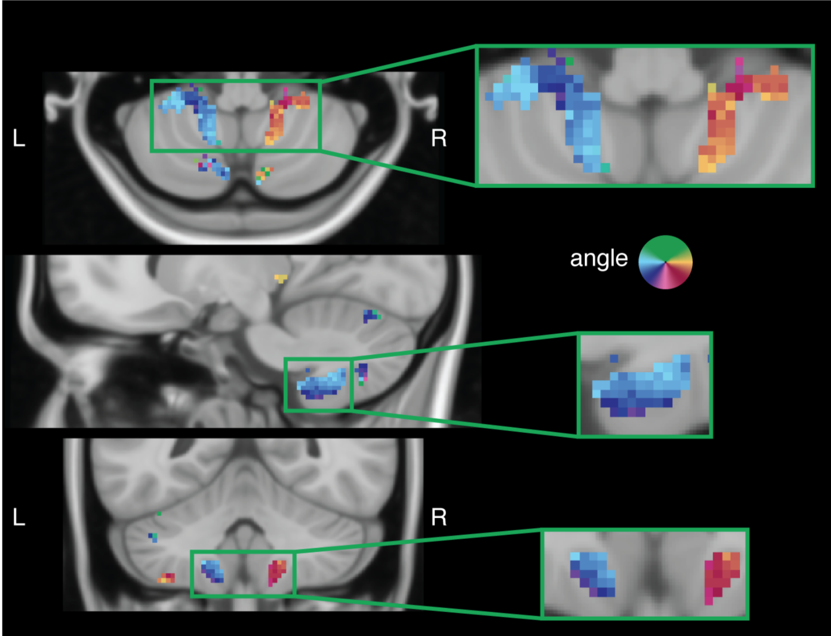
<p><img class="col two right" src="/img/posts/cerebellum_retmap.png" alt="Axial, sagittal and coronal slices of the cerebellum with voxels colored by preferred polar angle, showing visual field maps in both hemispheres." /></p>

<p><strong>New preprint on Biorxiv:</strong> While the cerebellum is instrumental for motor control, it is not traditionally implicated in vision. Here, we report the existence of 5 ipsilateral visual field maps in the human cerebellum. These maps are located within the oculomotor vermis and cerebellar nodes of the dorsal attention and visual networks. These findings imply that the cerebellum is closely involved in visuospatial cognition, and that its contributions are anchored in sensory coordinates.</p>]]></content><author><name>Tomas Knapen</name></author><summary type="html"><![CDATA[Retinotopic maps of visual space in the human cerebellum]]></summary><media:thumbnail xmlns:media="http://search.yahoo.com/mrss/" url="https://tknapen.github.io/assets/img/social-card.png" /><media:content medium="image" url="https://tknapen.github.io/assets/img/social-card.png" xmlns:media="http://search.yahoo.com/mrss/" /></entry></feed>