Topographic maps
How maps of the sensory world and of the body organize processing throughout the brain.
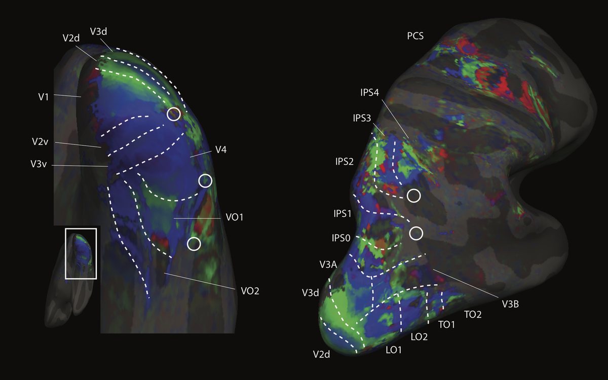
Early sensory cortex is laid out as maps: neighboring neurons respond to neighboring points on the retina or the skin. We ask how far this organization reaches beyond the classical sensory areas, how it changes with cognitive state and with eye movements, and what maps contribute to memory, to action and to perceiving other people’s bodies.
Approach
We estimate each brain location’s preferred position in visual space with population receptive field (pRF) models. Topographic connectivity derives preferred positions in visual space or on the body from the pattern of activity on a source map, such as primary visual cortex (V1) or primary somatosensory cortex (S1). It needs only brain activity, so it also works during movie watching and rest. We analyze large public datasets, such as the Human Connectome Project (HCP) retinotopy data, alongside individuals scanned over many runs.
What we have found
The cerebellum contains maps of visual space. In the HCP retinotopy data we found five ipsilateral retinotopic maps in three clusters of the human cerebellum (oculomotor vermis, lobule VIIb and lobule VIIIb), and released an atlas of them (van Es et al., 2019). Connectivity with V1 later revealed an eccentricity gradient in the oculomotor vermis during movie watching, absent in a simple mapping experiment, and a new topographic area in Crus II that appeared during movies but not at rest (Zuiderbaan et al., 2026).
Visual coding extends into the default network and hippocampus. In the human brain, the default network deactivates selectively depending on where a visual stimulus appears: it encodes spatial vision through deactivation (Szinte and Knapen, 2020). Connectivity with V1 revealed stable visual organization in the default-mode network and hippocampus, warped by cognitive state (Knapen, 2021). In the human hippocampus, pRF mapping showed strong biases for the contralateral visual field (Silson et al., 2021).
Body maps in visual cortex. A model that maps body-part tuning and visual field tuning at once revealed an array of somatotopic body maps across the human dorsolateral visual system during video watching. Their body-position tuning aligns with their visual tuning and predicts both visual field preferences and preferences for seeing particular body parts (Hedger et al., 2025).
Maps across eye movements. Motion and tilt aftereffects are tied to retinal position, not to world- or head-centered position (Knapen et al., 2009; Knapen et al., 2010). Around a saccade, information about its target reaches foveal retinotopic cortex. In human fMRI, activity matching the target’s orientation and size was remapped to the fovea, but not when observers only attended the target (Knapen et al., 2016), and peripheral targets could be decoded from foveal cortex without foveal stimulation (Kämmer et al., 2026).
Open questions
With colleagues, we have proposed that visuospatial coding gives cognition a common reference frame (Groen et al., 2021). What do maps in memory and default-network regions compute, and how do they reorganize during free viewing? How do aligned visual and bodily maps support action and social perception, and how similar is this organization across primate species?
Papers
13 papers on this theme, selected papers first. Filter the full list.
-
Vicarious body maps bridge vision and touch in the human brain
-
Visuospatial coding as ubiquitous scaffolding for human cognition
-
Topographic connectivity reveals task-dependent retinotopic processing throughout the human brain
-
Topographic Maps of Visual Space in the Human Cerebellum
-
Feedback of peripheral saccade targets to early foveal cortex
-
Task-Dependent Visual Topographic Connectivity in the Human Cerebellum
-
Representation of Contralateral Visual Space in the Human Hippocampus
-
Visual Organization of the Default Network
-
How Visual Cortical Organization Is Altered by Ophthalmologic and Neurologic Disorders
-
Oculomotor Remapping of Visual Information to Foveal Retinotopic Cortex
People
Xaver Funk
Postdoctoral researcher (from 2027)
Former lab members who worked on this theme: Daan van Es, Martin Szinte, Nick Hedger, Inês Veríssimo.
Software & data
prfmodel (Popylar)
BetaPython package for fitting population receptive field models (including compressive spatial summation, difference-of-Gaussians and divisive normalization variants) and related models such as connective field and contrast sensitivity models, on GPU-accelerated backends (TensorFlow, PyTorch or JAX, through Keras 3). Developed by the Popylar team with the Netherlands eScience Center.
pip install "prfmodel[tensorflow]"prfpy
AvailablePython package to simulate and fit population receptive field (pRF) models to time series data, including the divisive normalization pRF model. prfmodel, developed in the Popylar project, builds on it.
Vicarious somatotopy analysis code
AvailableAnalysis code for the vicarious body maps study, which fits connective field models of somatosensory and visual topography to 7T Human Connectome Project data.
Vicarious body-map atlas
AvailableAn atlas of the somatotopic body maps described in the vicarious body maps study. The paper's data availability statement describes how to obtain it.
Cerebellar retinotopy atlas
AvailableAtlas of the retinotopic maps in the human cerebellum (oculomotor vermis, lobule VIIb and lobule VIIIb), derived from Human Connectome Project retinotopy data, in 2 mm MNI space, with the code that produced it.